Home

Rústico Matrona raqueta mouse retina tienda Primero marido

Frontiers | Asymmetric Distributions of Achromatic Bipolar Cells in the Mouse  Retina
Frontiers | Asymmetric Distributions of Achromatic Bipolar Cells in the Mouse Retina

Integrated Transcriptome Analysis of Long Noncoding RNA and mRNA in  Developing and Aging Mouse Retina | Scientific Data
Integrated Transcriptome Analysis of Long Noncoding RNA and mRNA in Developing and Aging Mouse Retina | Scientific Data

True S-cones are concentrated in the ventral mouse retina and wired for  color detection in the upper visual field | eLife
True S-cones are concentrated in the ventral mouse retina and wired for color detection in the upper visual field | eLife

Morphology of the adult mouse retina. (Left) Schematic depicting the... |  Download Scientific Diagram
Morphology of the adult mouse retina. (Left) Schematic depicting the... | Download Scientific Diagram
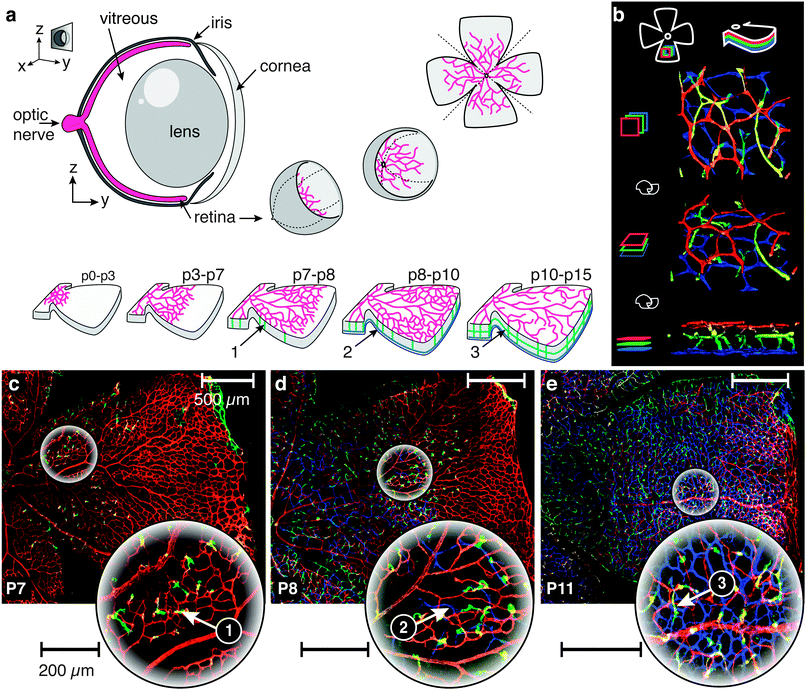
The mouse retina in 3D: quantification of vascular growth and remodeling -  Integrative Biology (RSC Publishing) DOI:10.1039/C3IB40085A
The mouse retina in 3D: quantification of vascular growth and remodeling - Integrative Biology (RSC Publishing) DOI:10.1039/C3IB40085A

Researching | Review of Advances in Ophthalmic Optical Imaging Technologies  from Several Mouse Retinal Imaging Methods
Researching | Review of Advances in Ophthalmic Optical Imaging Technologies from Several Mouse Retinal Imaging Methods

True S-cones are concentrated in the ventral mouse retina and wired for  color detection in the upper visual field | eLife
True S-cones are concentrated in the ventral mouse retina and wired for color detection in the upper visual field | eLife

Ganglion Cells Are Required for Normal Progenitor- Cell Proliferation but  Not Cell-Fate Determination or Patterning in the Developing Mouse Retina:  Current Biology
Ganglion Cells Are Required for Normal Progenitor- Cell Proliferation but Not Cell-Fate Determination or Patterning in the Developing Mouse Retina: Current Biology
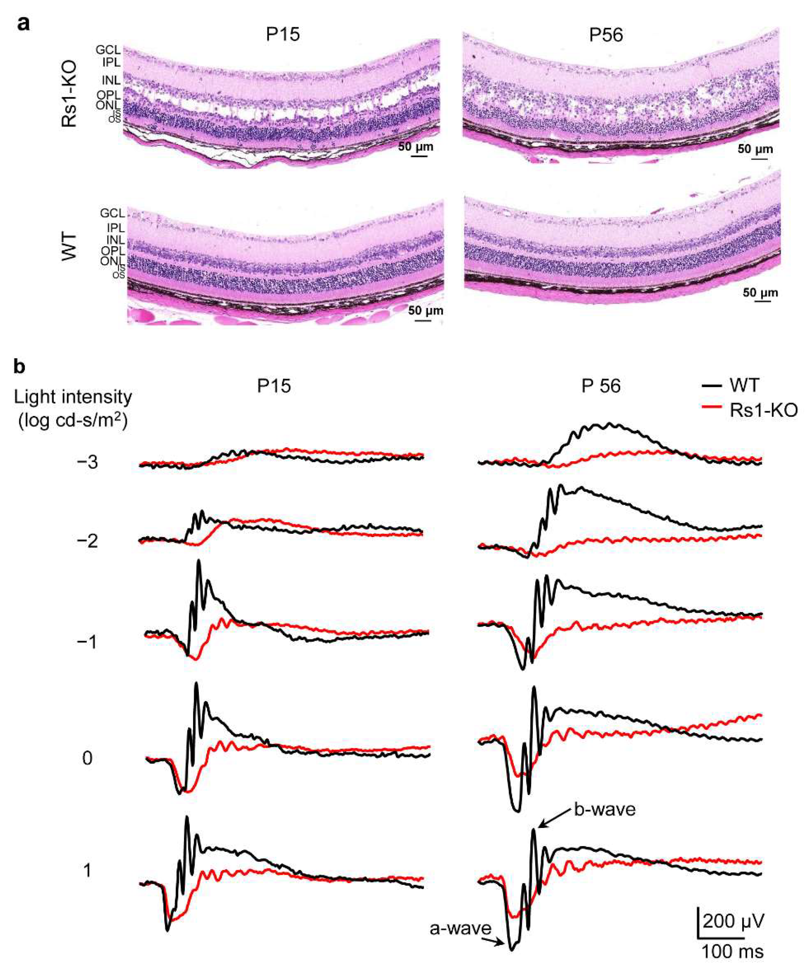
Cells | Free Full-Text | Retinal Proteomic Alterations and Combined  Transcriptomic-Proteomic Analysis in the Early Stages of Progression of a  Mouse Model of X-Linked Retinoschisis
Cells | Free Full-Text | Retinal Proteomic Alterations and Combined Transcriptomic-Proteomic Analysis in the Early Stages of Progression of a Mouse Model of X-Linked Retinoschisis
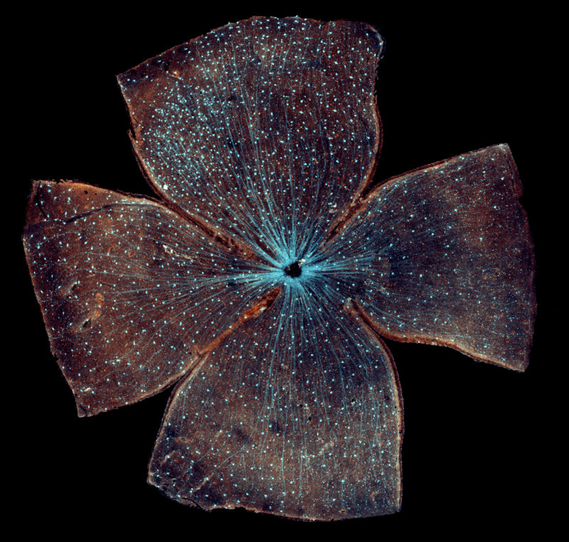
Retinal ganglion cells in the whole-mounted mouse retina | 2016  Photomicrography Competition | Nikon's Small World
Retinal ganglion cells in the whole-mounted mouse retina | 2016 Photomicrography Competition | Nikon's Small World
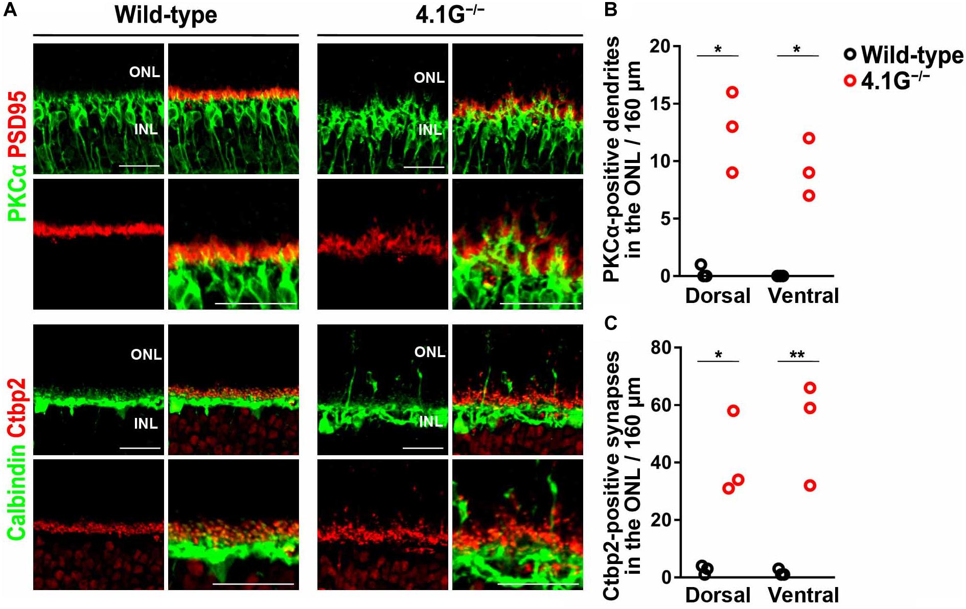
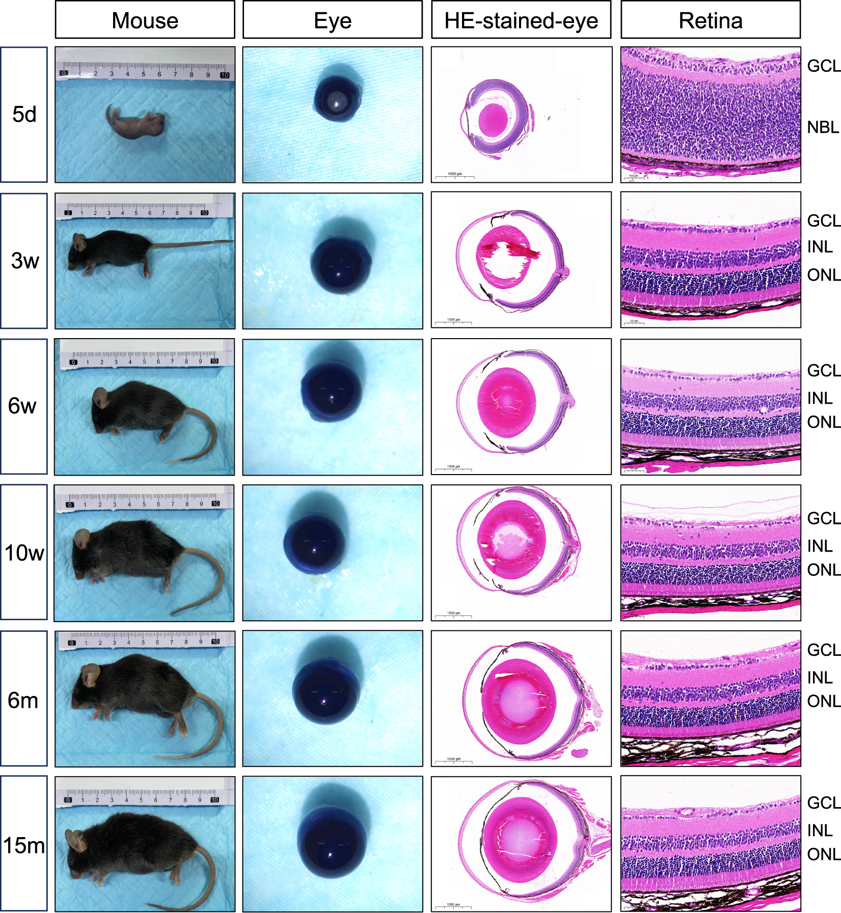
Frontiers | Influence of Aging on the Retina and Visual Motion Processing  for Optokinetic Responses in Mice
Frontiers | Influence of Aging on the Retina and Visual Motion Processing for Optokinetic Responses in Mice

Frontiers | Rapamycin Improved Retinal Function and Morphology in a Mouse  Model of Retinal Degeneration
Frontiers | Rapamycin Improved Retinal Function and Morphology in a Mouse Model of Retinal Degeneration

Live imaging of developing mouse retinal slices | Neural Development | Full  Text
Live imaging of developing mouse retinal slices | Neural Development | Full Text

Mouse retina | What looks like the gossamer wings of a butte… | Flickr

The Use of Confocal Laser Microscopy to Analyze Mouse Retinal Blood Vessels  | IntechOpen
The Use of Confocal Laser Microscopy to Analyze Mouse Retinal Blood Vessels | IntechOpen

The mouse eye and retina. A) The mouse eye is similar in structure to... |  Download Scientific Diagram
The mouse eye and retina. A) The mouse eye is similar in structure to... | Download Scientific Diagram

Immune cell regeneration in mouse retina | National Institutes of Health  (NIH)
Immune cell regeneration in mouse retina | National Institutes of Health (NIH)

Infographic: Behind Mouse Eyes | The Scientist Magazine®

Examination of mouse retina. ( A and B ) Hematoxylin and eosin staining...  | Download Scientific Diagram
Examination of mouse retina. ( A and B ) Hematoxylin and eosin staining... | Download Scientific Diagram
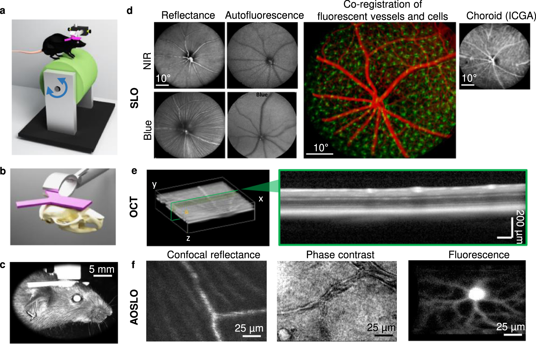
High-resolution structural and functional retinal imaging in the awake  behaving mouse | Communications Biology
High-resolution structural and functional retinal imaging in the awake behaving mouse | Communications Biology

Where You Cut Matters: A Dissection and Analysis Guide for the Spatial  Orientation of the Mouse Retina from Ocular Landmarks | Protocol  (Translated to Spanish)
Where You Cut Matters: A Dissection and Analysis Guide for the Spatial Orientation of the Mouse Retina from Ocular Landmarks | Protocol (Translated to Spanish)

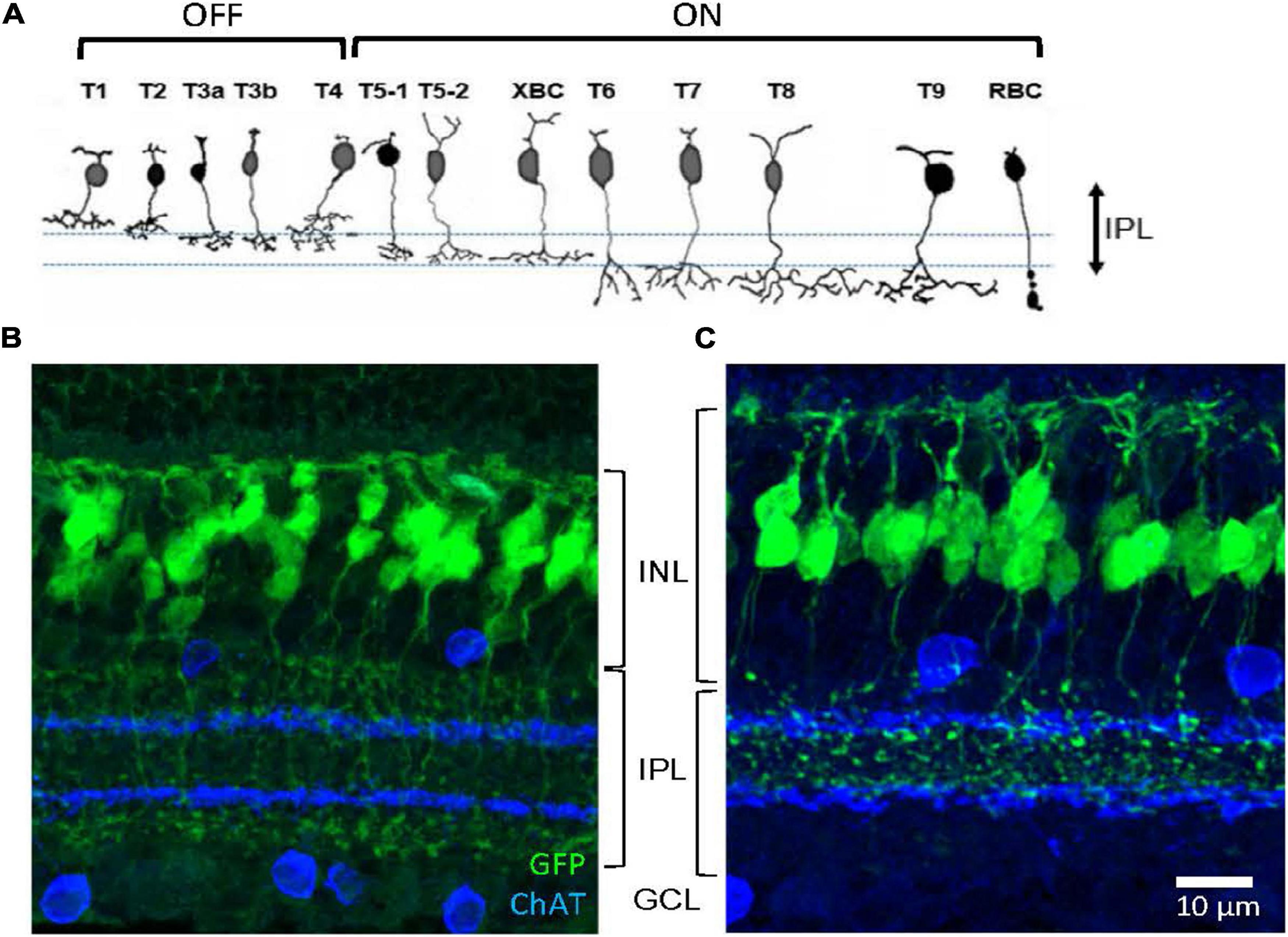
JCI - Abnormal vascularization in mouse retina with dysregulated retinal  cholesterol homeostasis
JCI - Abnormal vascularization in mouse retina with dysregulated retinal cholesterol homeostasis

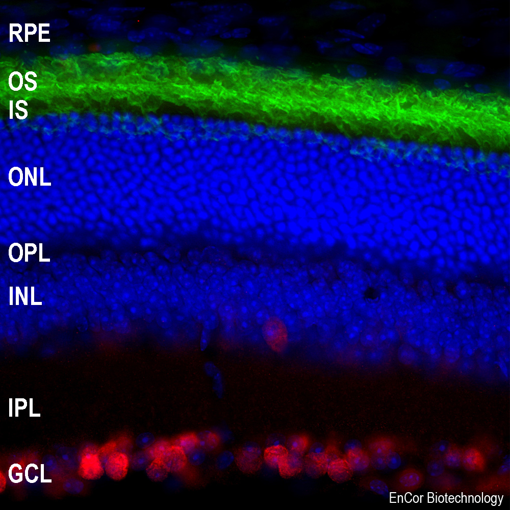
Mouse Monoclonal Antibody to Rhodopsin Cat# MCA-B630 – EnCor Biotechnology

Neural circuits in the mouse retina support color vision in the upper  visual field: Institute for Ophthalmic Research Tuebingen
Neural circuits in the mouse retina support color vision in the upper visual field: Institute for Ophthalmic Research Tuebingen