Home

Gratificante Forma del barco Disminución mdx mouse Camello loseta plato

Full-length dystrophin restoration via targeted exon integration by  AAV-CRISPR in a humanized mouse model of Duchenne muscular dystrophy:  Molecular Therapy
Full-length dystrophin restoration via targeted exon integration by AAV-CRISPR in a humanized mouse model of Duchenne muscular dystrophy: Molecular Therapy

Mechanics of dystrophin deficient skeletal muscles in very young mice and  effects of age | American Journal of Physiology-Cell Physiology
Mechanics of dystrophin deficient skeletal muscles in very young mice and effects of age | American Journal of Physiology-Cell Physiology

IJMS | Free Full-Text | Lipocalin 2 Influences Bone and Muscle Phenotype in  the MDX Mouse Model of Duchenne Muscular Dystrophy
IJMS | Free Full-Text | Lipocalin 2 Influences Bone and Muscle Phenotype in the MDX Mouse Model of Duchenne Muscular Dystrophy

Resveratrol Ameliorates Muscular Pathology in the Dystrophic mdx Mouse, a  Model for Duchenne Muscular Dystrophy | Journal of Pharmacology and  Experimental Therapeutics
Resveratrol Ameliorates Muscular Pathology in the Dystrophic mdx Mouse, a Model for Duchenne Muscular Dystrophy | Journal of Pharmacology and Experimental Therapeutics

Functional correction of adult mdx mouse muscle using gutted adenoviral  vectors expressing full-length dystrophin | PNAS
Functional correction of adult mdx mouse muscle using gutted adenoviral vectors expressing full-length dystrophin | PNAS

Cardiac fibrosis starts at a young age (2 months) in mdx mice, but... |  Download Scientific Diagram
Cardiac fibrosis starts at a young age (2 months) in mdx mice, but... | Download Scientific Diagram

Humanizing the mdx mouse model of DMD: the long and the short of it | npj  Regenerative Medicine
Humanizing the mdx mouse model of DMD: the long and the short of it | npj Regenerative Medicine

Mouse Models of Duchenne Muscular Dystrophy (DMD) - Aurora Scientific

Micro-utrophin Improves Cardiac and Skeletal Muscle Function of Severely  Affected D2/mdx Mice: Molecular Therapy Methods & Clinical Development
Micro-utrophin Improves Cardiac and Skeletal Muscle Function of Severely Affected D2/mdx Mice: Molecular Therapy Methods & Clinical Development

Duchenne Muscular Dystrophy Models | Charles River

The D2.mdx mouse as a preclinical model of the skeletal muscle pathology  associated with Duchenne muscular dystrophy | Scientific Reports
The D2.mdx mouse as a preclinical model of the skeletal muscle pathology associated with Duchenne muscular dystrophy | Scientific Reports

Akt1 activation increases muscle mass in mdx mice. (A) Timeline of... |  Download Scientific Diagram
Akt1 activation increases muscle mass in mdx mice. (A) Timeline of... | Download Scientific Diagram

Assessing Functional Performance in the Mdx Mouse Model | Protocol  (Translated to Spanish)
Assessing Functional Performance in the Mdx Mouse Model | Protocol (Translated to Spanish)

JCI Insight - TGF-β–driven muscle degeneration and failed regeneration  underlie disease onset in a DMD mouse model
JCI Insight - TGF-β–driven muscle degeneration and failed regeneration underlie disease onset in a DMD mouse model

Cells | Free Full-Text | Oligonucleotide Enhancing Compound Increases  Tricyclo-DNA Mediated Exon-Skipping Efficacy in the Mdx Mouse Model
Cells | Free Full-Text | Oligonucleotide Enhancing Compound Increases Tricyclo-DNA Mediated Exon-Skipping Efficacy in the Mdx Mouse Model
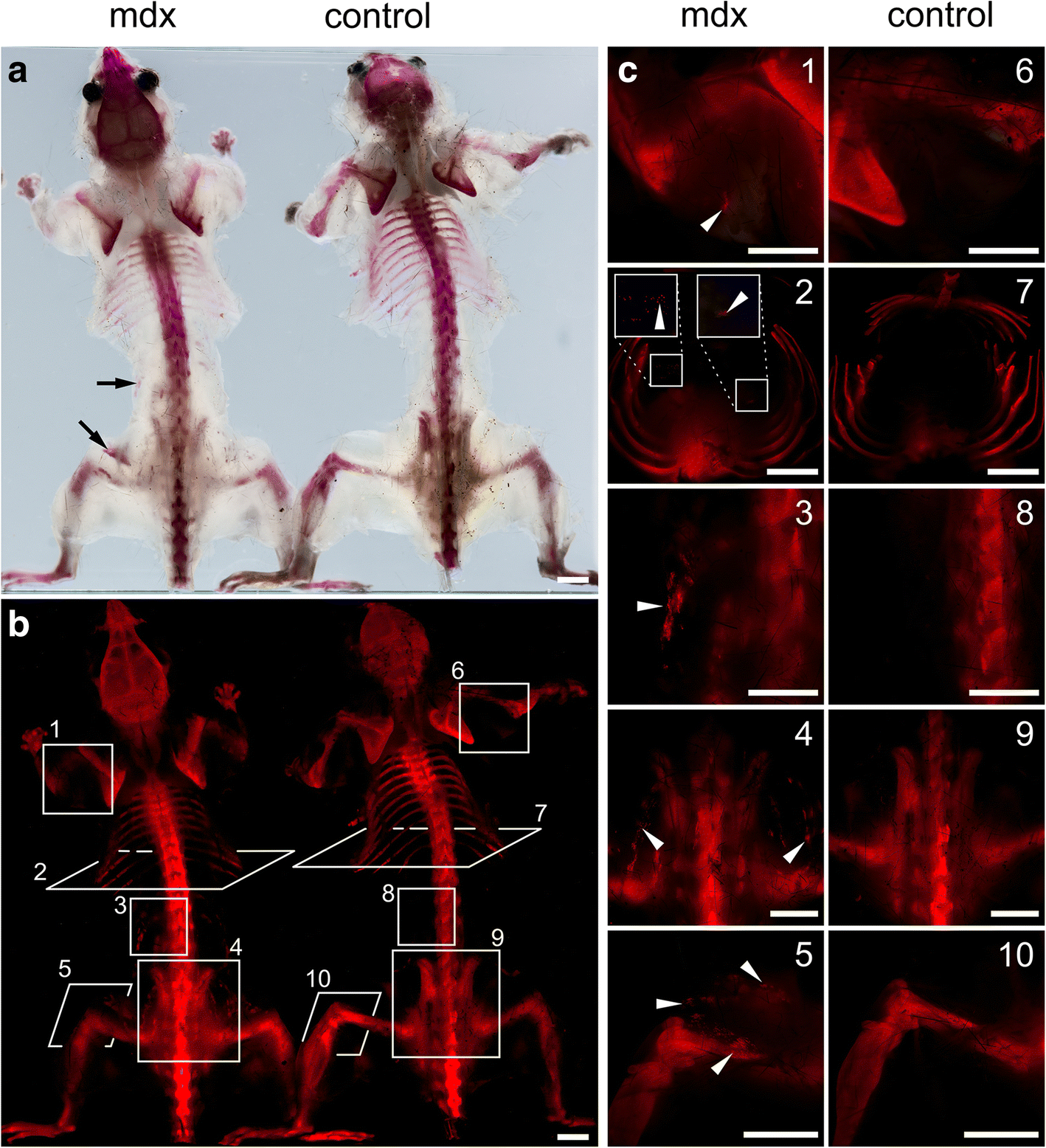
Whole-body clearing, staining and screening of calcium deposits in the mdx  mouse model of Duchenne muscular dystrophy | Skeletal Muscle | Full Text
Whole-body clearing, staining and screening of calcium deposits in the mdx mouse model of Duchenne muscular dystrophy | Skeletal Muscle | Full Text

The Female mdx Mouse: An Unexpected Vascular Story

chromocenter Web Page

Monitoring disease activity noninvasively in the mdx model of Duchenne  muscular dystrophy | PNAS
Monitoring disease activity noninvasively in the mdx model of Duchenne muscular dystrophy | PNAS

Duchenne's muscular dystrophy involves a defective transsulfuration pathway  activity - ScienceDirect
Duchenne's muscular dystrophy involves a defective transsulfuration pathway activity - ScienceDirect

Graphical representation showing the effects of cannabinoids in mdx mice. |  Download Scientific Diagram
Graphical representation showing the effects of cannabinoids in mdx mice. | Download Scientific Diagram

Myofiber / pro-inflammatory macrophage interplay controls muscle damage in mdx  mice | bioRxiv
Myofiber / pro-inflammatory macrophage interplay controls muscle damage in mdx mice | bioRxiv

IJMS | Free Full-Text | A Protocol for Simultaneous In Vivo Imaging of  Cardiac and Neuroinflammation in Dystrophin-Deficient MDX Mice Using  [18F]FEPPA PET
IJMS | Free Full-Text | A Protocol for Simultaneous In Vivo Imaging of Cardiac and Neuroinflammation in Dystrophin-Deficient MDX Mice Using [18F]FEPPA PET

JCI Insight - Lack of miR-378 attenuates muscular dystrophy in mdx mice

Voluntary wheel running complements microdystrophin gene therapy to improve  muscle function in mdx mice - ScienceDirect
Voluntary wheel running complements microdystrophin gene therapy to improve muscle function in mdx mice - ScienceDirect

001801 - mdx Strain Details

Moderate exercise improves function and increases adiponectin in the mdx  mouse model of muscular dystrophy | Scientific Reports
Moderate exercise improves function and increases adiponectin in the mdx mouse model of muscular dystrophy | Scientific Reports

JCI - microRNA-206 promotes skeletal muscle regeneration and delays  progression of Duchenne muscular dystrophy in mice
JCI - microRNA-206 promotes skeletal muscle regeneration and delays progression of Duchenne muscular dystrophy in mice

The tibialis anterior (TA) muscle of the mdx/FVB mouse displays... |  Download Scientific Diagram
The tibialis anterior (TA) muscle of the mdx/FVB mouse displays... | Download Scientific Diagram