Home

difícil Psicológico En el piso coronal mouse brain Viva cerca más y más

Quantitative Neuroanatomical Phenotyping of the Embryonic Mouse Brain -  Nguyen - 2022 - Current Protocols - Wiley Online Library
Quantitative Neuroanatomical Phenotyping of the Embryonic Mouse Brain - Nguyen - 2022 - Current Protocols - Wiley Online Library
File:201510 Mouse brain coronal section.png - Wikimedia Commons

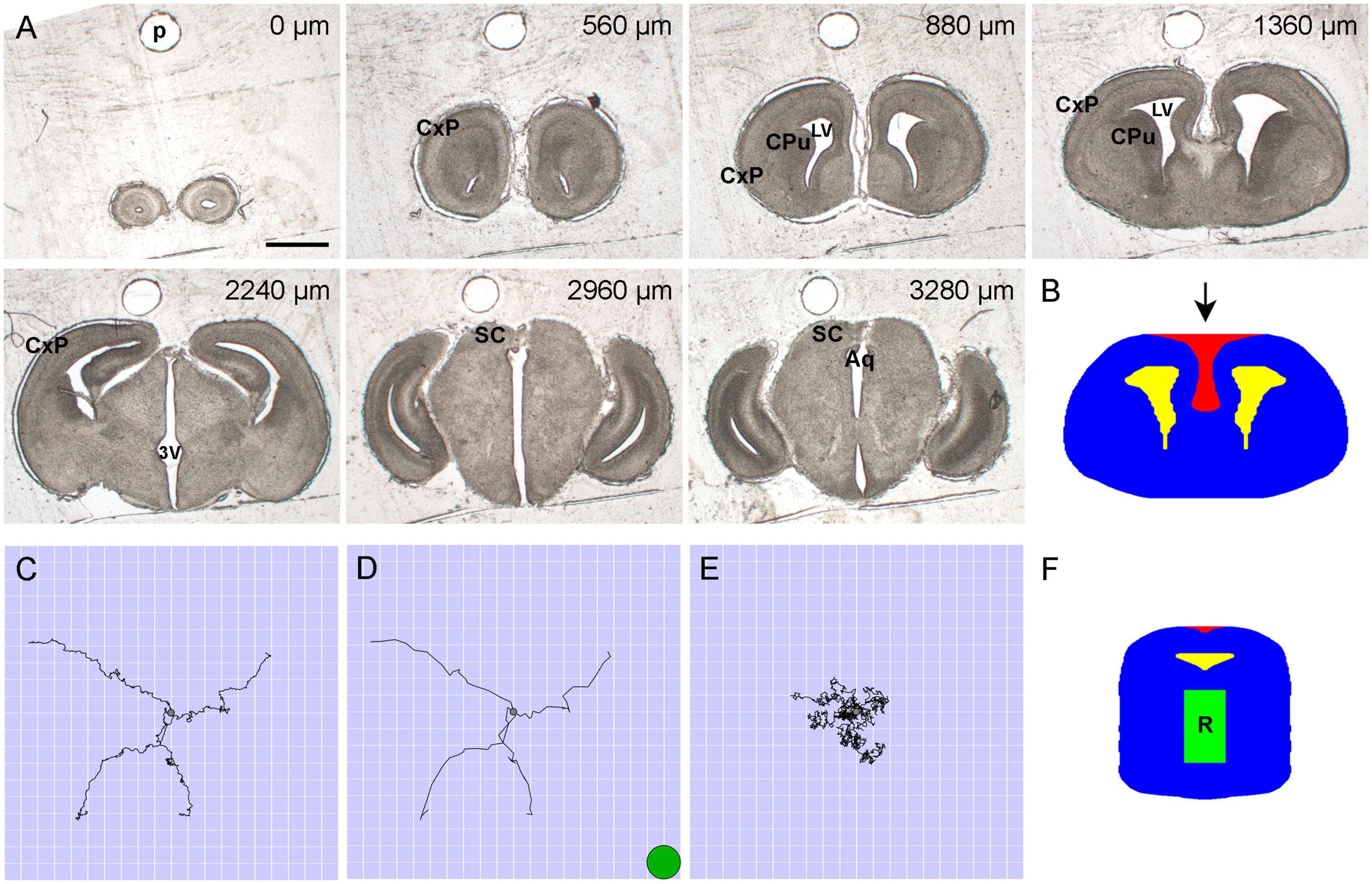
Frontiers | Predicting the distribution of serotonergic axons: a  supercomputing simulation of reflected fractional Brownian motion in a 3D-mouse  brain model
Frontiers | Predicting the distribution of serotonergic axons: a supercomputing simulation of reflected fractional Brownian motion in a 3D-mouse brain model

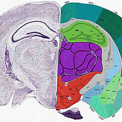
Atlas Thumbnails :: Allen Brain Atlas: Mouse Brain

Mouse Brain, illustration - Stock Image - C030/6193 - Science Photo Library

Coronal section of a mouse brain showing the SVZ for harvesting NSCs.... |  Download Scientific Diagram
Coronal section of a mouse brain showing the SVZ for harvesting NSCs.... | Download Scientific Diagram

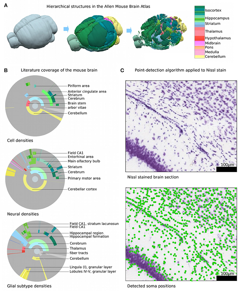
Frontiers | A Cell Atlas for the Mouse Brain

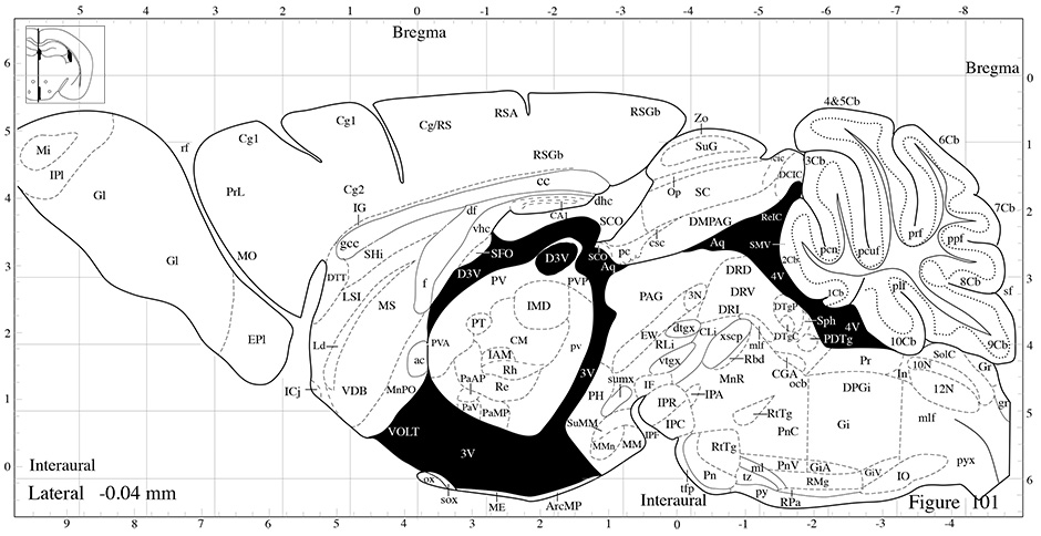
The Mouse Brain in Stereotaxic Coordinates: Compact Second Edition:  9780125476379: Medicine & Health Science Books @ Amazon.com
The Mouse Brain in Stereotaxic Coordinates: Compact Second Edition: 9780125476379: Medicine & Health Science Books @ Amazon.com

Scientists create atlas of mouse brain regions down to single cells

High Resolution Mouse Brain Atlas - Methods

A method to estimate the cellular composition of the mouse brain from  heterogeneous datasets | bioRxiv
A method to estimate the cellular composition of the mouse brain from heterogeneous datasets | bioRxiv

High Resolution Mouse Brain Atlas

Coronal section of the mouse brain illustrating the main areas of the... |  Download Scientific Diagram
Coronal section of the mouse brain illustrating the main areas of the... | Download Scientific Diagram
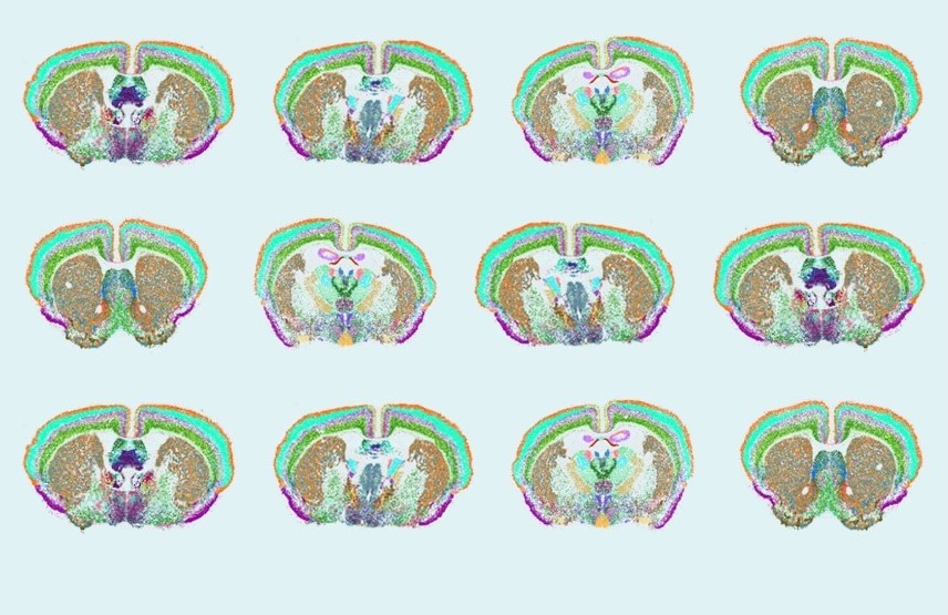
Scientists unveil complete cell map of a whole mammalian brain | National  Institutes of Health (NIH)
Scientists unveil complete cell map of a whole mammalian brain | National Institutes of Health (NIH)
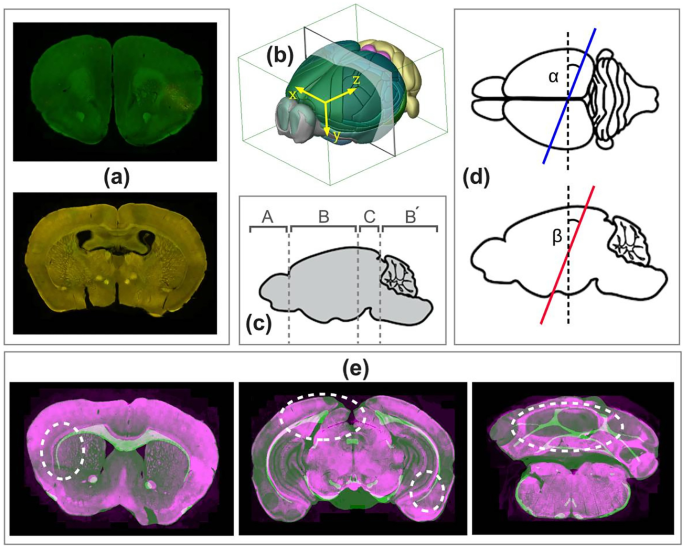
Sección coronal de cerebro de ratón cp 1

Localization and Registration of 2D Histological Mouse Brain Images in 3D  Atlas Space | Neuroinformatics
Localization and Registration of 2D Histological Mouse Brain Images in 3D Atlas Space | Neuroinformatics

Free Mouse brain (coronal, lateral ventricles) Icons, Symbols & Images |  BioRender
Free Mouse brain (coronal, lateral ventricles) Icons, Symbols & Images | BioRender

FMRP in Mouse brain | College of Medicine

Mouse Brain Anatomy Coronal Sections

Geometry of the coronal and sagittal sections of a mouse brain used... |  Download Scientific Diagram
Geometry of the coronal and sagittal sections of a mouse brain used... | Download Scientific Diagram
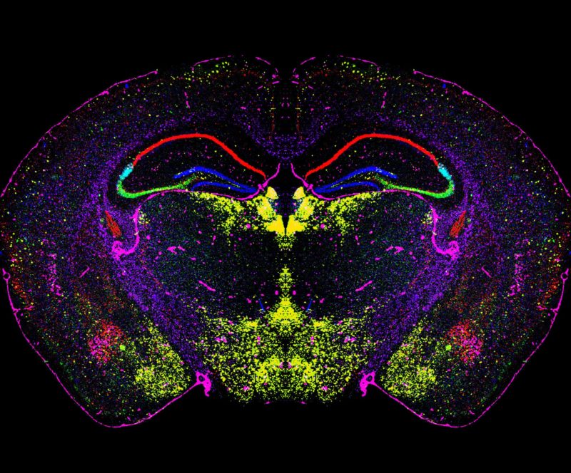
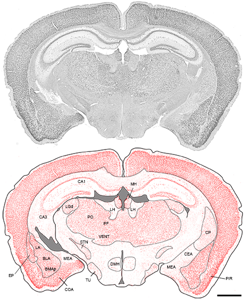
Mouse Brain Atlas
Mouse Brain Atlas

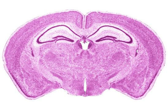
Coronal sections of a 10 week old mouse brain | 2004 Photomicrography  Competition | Nikon's Small World
Coronal sections of a 10 week old mouse brain | 2004 Photomicrography Competition | Nikon's Small World

A) Representative coronal sections of a mouse brain at levels R265,... |  Download Scientific Diagram
A) Representative coronal sections of a mouse brain at levels R265,... | Download Scientific Diagram

Mouse C57 Brain Coronal Frozen Sections – Zyagen

IJMS | Free Full-Text | Phosphorylation of Histone H2AX in the Mouse Brain  from Development to Senescence
IJMS | Free Full-Text | Phosphorylation of Histone H2AX in the Mouse Brain from Development to Senescence

Schematic diagrams of coronal sections of mice brain illustrating where  FosB/ΔFosB immunoreactivity was quantified in the following regions:  prelimbic cortex (PL), infralimbic cortex (IL), motor cortex (M1/2),  nucleus accumbens core (NAcC), nucleus
Schematic diagrams of coronal sections of mice brain illustrating where FosB/ΔFosB immunoreactivity was quantified in the following regions: prelimbic cortex (PL), infralimbic cortex (IL), motor cortex (M1/2), nucleus accumbens core (NAcC), nucleus

BrainInfo
BrainInfo

Prenatal Mouse Brain Atlas: Color images and annotated diagrams of:  Gestational Days 12, 14, 16 and 18 Sagittal, coronal and horizontal section  : Amazon.es: Libros
Prenatal Mouse Brain Atlas: Color images and annotated diagrams of: Gestational Days 12, 14, 16 and 18 Sagittal, coronal and horizontal section : Amazon.es: Libros